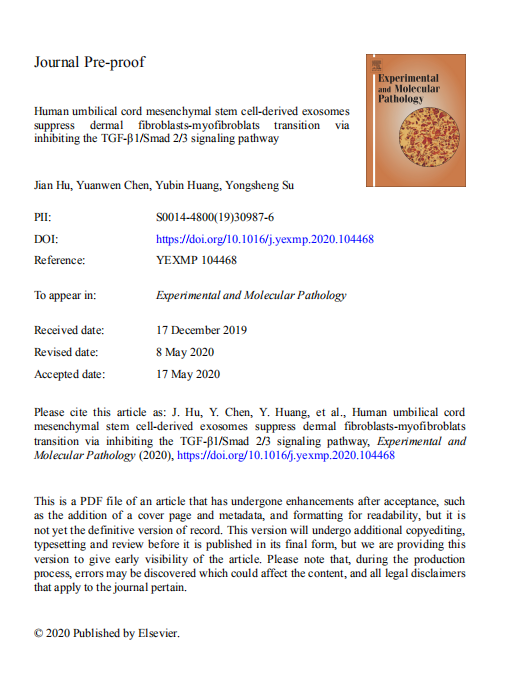
【文獻(xiàn)標(biāo)題】Human umbilical cord mesenchymal stem cell-derived exosomes suppress dermal fibroblasts-myofibroblats transition via inhibiting the TGF-β1/Smad 2/3 signaling pathway
【作者】Jian Hu, Yuanwen Chen, Yubin Huang,et.al
【作者單位】深圳市寶安區(qū)人民醫(yī)院(The People’s Hospital of Bao’an Shenzhen)
【文獻(xiàn)中引用產(chǎn)品】
【關(guān)鍵詞】Human umbilical cord mesenchymal stem cells�,Exosome, TGF-β1/Smad2/3 signaling pathway��,Dermal ibroblasts Myofibroblast
【DOI】https://doi.org/10.1016/j.yexmp.2020.104468
【影響因子(IF)】2.39
【出版期刊】《Experimental and Molecular Pathology》
【產(chǎn)品原文引用】
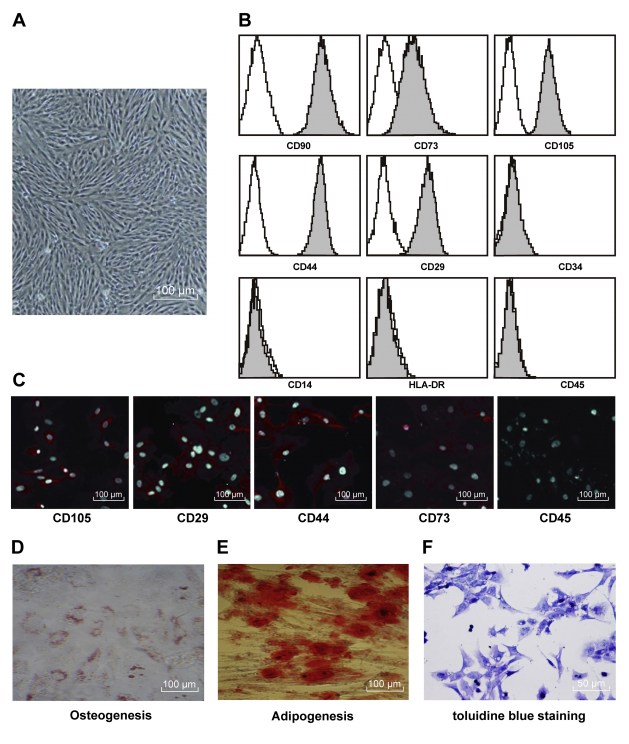
The hUC-MSCs or fibroblasts in exponential phase were seeded at 1 × 10⁵ cells/mL and 3 mL/well in a 6-well plate with a cover glass. After culture for 24 h at 37℃ with 5% CO2, culture medium was discarded, and cells were fixed with paraformaldehyde (Shanghai HengYuan Biological Technology Co., Ltd., Shanghai, China) for 10 min. After discarding the fixative solution, the cover glasses were rinsed by PBS 3 times, each for 3 min. PBS was then absorbed with absorbent papers, normal goat serum was dripped on the glass, and fibroblasts were sealed for 30min at room temperature. The sealing liquid was absorbed by absorbent papers without washes. After that, diluted primary antibodies (supplementary Table 1) were dripped into each cover glass, which was incubated in a wet box at 4℃ overnight. Afterwards, the fluorescent secondary antibody goat anti-rat IgG H&L (Alexa Fluor® 488) (1/200, ab150113) was added. Then the glass was dripped with 4',6-diamidino-2-phenylindole (DAPI) for nucleus staining, and sealed with sealing liquid containing anti-fluorescent quenching agent, and then observed under the fluorescence microscope. The above antibodies were all purchased from Abcam Inc. (Cambridge, MA, USA).
完整版PDF文獻(xiàn)請咨詢在線客服或者電話聯(lián)系我司業(yè)務(wù)員�!
更多公司福利請關(guān)注“恒遠(yuǎn)生物”微信公眾號���!


